What is Oxytocin?
Oxytocin is a nonapeptide hormone synthesized in the supraoptic and paraventricular nuclei of the hypothalamus and released through the posterior pituitary gland. Discovered in the early 20th century and traditionally associated with reproductive functions including parturition and lactation, research over the past two decades has revealed oxytocin's extensive roles in metabolic regulation, tissue regeneration, and cardiovascular homeostasis.
The peptide exerts its effects by binding to oxytocin receptors (OTRs), which are G-protein-coupled receptors expressed throughout the body including skeletal muscle tissue, bone marrow, cardiac tissue, pancreatic islets, adipose tissue, and vascular endothelium. This widespread receptor distribution underlies oxytocin's diverse physiological effects on tissue maintenance, metabolic function, and cellular protection.
Oxytocin levels naturally decline with age, with circulating concentrations decreasing significantly in individuals over 70 compared to younger adults. This age-related decline correlates with reduced muscle regenerative capacity, increased adiposity, decreased bone mineral density, and compromised metabolic function. The decline in endogenous oxytocin is now recognized as a contributing factor to sarcopenia, osteoporosis, metabolic syndrome, and cardiovascular disease associated with aging.
The hormone's mechanism of action involves multiple signaling pathways including MAPK/ERK activation for muscle stem cell proliferation, AMPK pathway stimulation for metabolic regulation, nitric oxide and atrial natriuretic peptide (ANP) production for cardiovascular effects, and modulation of inflammatory cytokines for tissue protection. Oxytocin also directly influences pancreatic function by stimulating insulin secretion and enhancing β-cell responsivity to glucose.
Notably, oxytocin represents the first FDA-approved molecule identified with anti-aging regenerative properties, as synthetic oxytocin (Pitocin) has been safely used in clinical practice for decades in obstetric applications. This established safety profile, combined with emerging evidence of its therapeutic potential in age-related tissue decline and metabolic disorders, positions oxytocin as a promising candidate for therapeutic applications beyond its traditional reproductive uses.
Oxytocin Structure
Chemical Structure
2D Structure
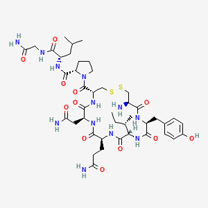
3D Structure

Chemical Properties
| CAS Number | 50-56-6 |
|---|---|
| Molecular Formula | C43H66N12O12S2 |
| Molecular Weight | 1007.2 g/mol |
| IUPAC Name |
(2S)-1-[(4R,7S,10S,13S,16S,19R)-19-amino-7-(2-amino-2-oxoethyl)-10-(3-amino-3-oxopropyl)-13-[(2S)-butan-2-yl]-16-[(4-hydroxyphenyl)methyl]-6,9,12,15,18-pentaoxo-1,2-dithia-5,8,11,14,17-pentazacycloicosane-4-carbonyl]-N-[(2S)-1-[(2-amino-2-oxoethyl)amino]-4-methyl-1-oxopentan-2-yl]pyrrolidine-2-carboxamide
|
| InChIKey | XNOPRXBHLZRZKH-DSZYJQQASA-N |
Oxytocin Research
Muscle Regeneration and Anti-Aging
Research demonstrates oxytocin plays a critical role in muscle maintenance and regeneration, with plasma oxytocin levels and muscle oxytocin receptor expression declining dramatically with age. Studies published in Nature Communications established that oxytocin is required for skeletal muscle tissue regeneration and homeostatic maintenance, with systemic oxytocin administration rapidly improving muscle regeneration in aged mice by enhancing aged muscle stem cell activation and proliferation through MAPK/ERK signaling pathway activation.
In controlled experiments, oxytocin treatment restored muscle repair in old mice to approximately 80% of the regenerative capacity observed in young mice, representing significant functional improvement. The peptide specifically targets muscle stem cells (satellite cells), which become reversibly inhibited in the aged niche but can be rescued for productive tissue repair through oxytocin intervention. Importantly, oxytocin administration to young mice produced no significant change in muscle regeneration, demonstrating the peptide selectively enhances aged tissue stem cells without causing uncontrolled cellular division.
Genetic studies using oxytocin knockout mice revealed that lack of oxytocin leads to premature sarcopenia, with mice deficient in the oxytocin gene displaying progressive decline in muscle regeneration noticeable at 3 months of age and pronounced at 12 months—comparable to the decline measured between young and old wild-type mice. This demonstrates oxytocin deficiency accelerates age-related muscle deterioration.
Human clinical trials have confirmed oxytocin's muscle-preserving effects. A randomized controlled trial of intranasal oxytocin administration (24 IU four times daily) for 8 weeks in older adults with sarcopenic obesity demonstrated significant improvements in lean muscle mass while reducing adiposity. The study showed oxytocin treatment increased lean body mass and lowered LDL cholesterol without adversely affecting muscle tissue, suggesting therapeutic potential for combating age-related muscle loss while promoting favorable body composition changes.
Sources:
- Elabd C, et al. "Oxytocin is an age-specific circulating hormone that is necessary for muscle maintenance and regeneration." Nature Communications. 2014;5:4082. https://www.nature.com/articles/ncomms5082
- Elabd C, et al. "Oxytocin is an age-specific circulating hormone that is necessary for muscle maintenance and regeneration." PMC. 2014. https://pmc.ncbi.nlm.nih.gov/articles/PMC4512838/
- Pinkerton JV, et al. "Intranasal Oxytocin Improves Lean Muscle Mass and Lowers LDL Cholesterol in Older Adults with Sarcopenic Obesity: A Pilot Randomized Controlled Trial." PMC. 2021. https://pmc.ncbi.nlm.nih.gov/articles/PMC8567747/
Metabolic Health and Fat Loss
Oxytocin demonstrates potent effects on energy metabolism and body composition through multiple mechanisms including enhanced lipolysis, increased fatty acid β-oxidation, improved metabolic flexibility, and stimulation of thermogenesis. Research published in PLOS ONE established that chronic central oxytocin infusion in diet-induced obese rats lowered body weight gain by more than 50% compared to controls, without changes in food intake, indicating improved metabolic efficiency rather than simple appetite suppression.
The peptide increases adipose tissue lipolysis and fatty acid β-oxidation through activation of PPAR-alpha signaling via production of oleoylethanolamide (OEA), a known endocannabinoid and PPAR-alpha ligand. Studies demonstrate oxytocin treatment significantly elevates expression of enzymes involved in lipolysis and fatty acid β-oxidation in epididymal adipose tissue, promoting utilization of stored fat as an energy source. Additionally, oxytocin stimulates brown adipocyte formation and thermogenesis in both brown adipose tissue (BAT) and skeletal muscle, contributing to increased energy expenditure.
Human clinical trials have confirmed oxytocin's metabolic benefits. An 8-week study of intranasal oxytocin (24 IU four times daily) in men and women with obesity demonstrated mean BMI reduction of 3.2 kg/m² with greater weight loss in subjects with higher degrees of obesity. Treatment also reduced waist and hip circumferences and improved lipid profiles with lower total cholesterol and LDL cholesterol levels. A single-dose study in men showed oxytocin administration resulted in decreased respiratory quotient within 30 minutes, indicating acute increase in fat oxidation as measured by indirect calorimetry.
Oxytocin administration in animal models demonstrates dose-dependent decreases in body weight gain, increased adipose tissue lipolysis and fatty acid β-oxidation, reduced glucose intolerance and insulin resistance, decreased visceral fat mass and adipocyte size, and amelioration of fatty liver. Studies show peripheral oxytocin treatment reduces visceral fat accumulation specifically, addressing the most metabolically harmful fat depot. Research indicates oxytocin's weight loss effects occur through both reduced caloric intake and increased energy expenditure, with the peptide promoting preferential preservation of lean body mass while reducing fat mass.
Sources:
- Deblon N, et al. "Mechanisms of the Anti-Obesity Effects of Oxytocin in Diet-Induced Obese Rats." PLOS ONE. 2011;6(9):e25565. https://journals.plos.org/plosone/article?id=10.1371/journal.pone.0025565
- Lawson EA, et al. "The effects of oxytocin on eating behaviour and metabolism in humans." PMC. 2018. https://pmc.ncbi.nlm.nih.gov/articles/PMC5868755/
- Zhang H, et al. "Treatment of Obesity and Diabetes Using Oxytocin or Analogs in Patients and Mouse Models." PLOS ONE. 2013;8(5):e61477. https://journals.plos.org/plosone/article?id=10.1371/journal.pone.0061477
- Ding C, et al. "Oxytocin in metabolic homeostasis: implications for obesity and diabetes management." Obesity Reviews. 2019;20(1):22-40. https://pmc.ncbi.nlm.nih.gov/articles/PMC7888317/
- Maejima Y, et al. "Peripheral oxytocin treatment ameliorates obesity by reducing food intake and visceral fat mass." Aging. 2011;3(12):1169-1177. https://www.aging-us.com/article/100408/text
- Yuan T, et al. "The effects of oxytocin to rectify metabolic dysfunction in obese mice are associated with increased thermogenesis." Acta Biochimica et Biophysica Sinica. 2020. https://www.sciencedirect.com/science/article/abs/pii/S0303720720302033
Bone Health and Mineralization
Oxytocin exerts anabolic effects on bone tissue by promoting osteoblast differentiation and function while modulating osteoclast activity, leading to increased bone formation and improved bone microarchitecture. Research demonstrates that both osteoblasts and osteoclasts express oxytocin receptors, and oxytocin directly controls differentiation of human mesenchymal stem cells toward osteogenic rather than adipogenic lineages.
Studies using oxytocin knockout mice show that animals lacking either oxytocin or its receptor develop low-turnover osteoporosis that worsens with age in both sexes. The skeletons of these mice display pronounced decreases in vertebral and femoral trabecular volume, with osteoblasts exhibiting lower mineralization activity and downregulation of all master genes for osteoblast differentiation. This establishes oxytocin as essential for maintaining skeletal homeostasis throughout life.
Oxytocin stimulates osteoblast mineralization by promoting oxytocin receptor translocation into the osteoblast nucleus and regulating the OPG/RANKL ratio through induction of intracytoplasmic calcium release and nitric oxide synthesis. While oxytocin increases osteoclast formation both directly through NFκB and MAPK signaling activation and indirectly through upregulation of RANK-L synthesis by osteoblasts, it simultaneously inhibits osteoclast resorptive function, resulting in net positive bone formation.
Human studies demonstrate significant associations between oxytocin levels and bone health. Research in postmenopausal women shows those with osteoporosis display significantly lower oxytocin levels (approximately 50 pg/mL) compared to healthy controls (110 pg/mL), with decreased oxytocin levels predictive of osteoporosis independent of age, estradiol, and leptin levels. A six-year prospective study found higher serum oxytocin levels associated with higher bone mineral density in postmenopausal women.
Animal studies confirm oxytocin administration prevents and reverses bone loss induced by estrogen deficiency. Systemic oxytocin treatment in ovariectomized mice (mimicking postmenopausal osteoporosis) prevented development of osteoporosis and reversed established bone loss by improving bone mineral density and microarchitecture. Studies show oxytocin's bone-protective effects are mediated through direct action on bone tissue rather than indirect hormonal mechanisms.
Sources:
- Elabd C, et al. "Oxytocin controls differentiation of human mesenchymal stem cells and reverses osteoporosis." Stem Cells. 2008;26(9):2399-2407. https://pubmed.ncbi.nlm.nih.gov/18599809/
- Colaianni G, et al. "The Oxytocin–Bone Axis." Journal of Neuroendocrinology. 2014;26(2):53-57. https://onlinelibrary.wiley.com/doi/10.1111/jne.12120
- Colaianni G, et al. "The Oxytocin-Bone Axis." PMC. 2014. https://pmc.ncbi.nlm.nih.gov/articles/PMC4108483/
- Breuil V, et al. "Oxytocin and Bone: Review and Perspectives." International Journal of Molecular Sciences. 2021;22(16):8551. https://pmc.ncbi.nlm.nih.gov/articles/PMC8395200/
- Sun L, et al. "Oxytocin and bone." American Journal of Physiology-Regulatory, Integrative and Comparative Physiology. 2014;307(6):R970-R977. https://journals.physiology.org/doi/full/10.1152/ajpregu.00040.2014
- Martins D, et al. "Oxytocin: From Biomarker to Therapy for Postmenopausal Osteoporosis." Women. 2025;5(3):27. https://www.mdpi.com/2673-4184/5/3/27
Glucose Homeostasis and Insulin Sensitivity
Oxytocin plays a significant role in glucose metabolism and insulin regulation through direct effects on pancreatic function and peripheral insulin sensitivity. Research demonstrates pancreatic islets harbor oxytocin receptors, and oxytocin administration stimulates insulin secretion from β-cells independent of glucose concentration through both central vagal cholinergic pathways and peripheral mechanisms involving phosphoinositide turnover and protein kinase C activation.
Clinical trials in healthy men published in Diabetes journal showed intranasal oxytocin administration significantly improved oral glucose tolerance and enhanced pancreatic β-cell responsivity. The study demonstrated oxytocin increased total β-cell responsivity markers and boosted dynamic insulin response to glucose, suggesting improved β-cell capacity to secrete insulin in response to rising glucose concentrations. Treatment resulted in more than twofold increase in insulin-dependent glucose tolerance and enhanced insulin sensitivity as indicated by stronger effects of insulin on stimulating glucose disposal and inhibiting hepatic glucose production.
Research in animal models demonstrates oxytocin-deficient mice display decreased insulin sensitivity and glucose intolerance, while oxytocin administration improves these parameters in both normal and obese diabetic mice. Studies show peripheral oxytocin administration improves insulin sensitivity and glucose tolerance in mice on standard and high-fat diets as well as in genetic models of diabetes (db/db mice). Treatment with oxytocin reduces fasting blood glucose levels and improves insulin sensitivity in leptin receptor-deficient mice over extended treatment periods.
Oxytocin appears to improve insulin sensitivity through multiple mechanisms including: reduction of glucotoxicity and lipotoxicity in tissues, modulation of cytokines such as leptin and adiponectin that influence insulin signaling, decreased visceral fat mass leading to reduced inflammatory mediators, and direct stimulation of glucose uptake in skeletal muscle cells. Studies demonstrate oxytocin triggers glucose uptake in young rat muscle cells and that oxytocin receptors in pancreatic Langerhans islets help regulate insulin and glucagon secretion.
Research indicates oxytocin provides protective effects on pancreatic β-cells by reducing endoplasmic reticulum stress and inflammation that contribute to β-cell dysfunction. In streptozotocin-induced diabetic models, oxytocin treatment significantly improved glucose tolerance and prevented impairment of first-phase and second-phase insulin secretion, demonstrating the peptide's capacity to protect β-cell function even under conditions of induced damage.
Sources:
- Klement J, et al. "Oxytocin Improves β-Cell Responsivity and Glucose Tolerance in Healthy Men." Diabetes. 2017;66(2):264-271. https://diabetesjournals.org/diabetes/article/66/2/264/35279/Oxytocin-Improves-Cell-Responsivity-and-Glucose
- Qian W, et al. "Two Birds with One Stone: Possible Dual-Role of Oxytocin in the Treatment of Diabetes and Osteoporosis." PMC. 2015. https://pmc.ncbi.nlm.nih.gov/articles/PMC4530313/
- Ding C, et al. "Oxytocin in metabolic homeostasis: implications for obesity and diabetes management." Obesity Reviews. 2019;20(1):22-40. https://onlinelibrary.wiley.com/doi/full/10.1111%2Fobr.12757
- Zhang H, et al. "Treatment of Obesity and Diabetes Using Oxytocin or Analogs in Patients and Mouse Models." PLOS ONE. 2013;8(5):e61477. https://journals.plos.org/plosone/article?id=10.1371/journal.pone.0061477
- Noble EE, et al. "Oxytocin as a Metabolic Modulator." IntechOpen. 2021. https://www.intechopen.com/chapters/76615
Cardiovascular Protection and Anti-Inflammatory Effects
Oxytocin demonstrates significant cardioprotective properties through multiple mechanisms including reduction of ischemia-reperfusion injury, anti-inflammatory effects, antioxidant activity, improved cardiac healing, and beneficial modulation of cardiovascular hemodynamics. Research shows acute oxytocin administration at the onset of reperfusion enhances cardiomyocyte viability and function by activating PI3K and Akt phosphorylation pathways along with downstream reperfusion injury salvage kinase (RISK) and signal transducer and activator of transcription (STAT) protein cardioprotective pathways.
Studies in experimentally induced myocardial infarction demonstrate continuous oxytocin delivery improves cardiac work, reduces apoptosis and inflammation, increases scar vascularization, and stimulates angiogenesis. Research published in Basic Research in Cardiology showed oxytocin treatment in rat myocardial infarction models significantly reduced inflammatory response, as evidenced by decreased expression of proinflammatory cytokines (IL-6, TNF-α, IL-1β) and reduced immune cell infiltration into damaged cardiac tissue.
Oxytocin's cardioprotective mechanisms involve production of cyclic GMP (cGMP) stimulated by local release of atrial natriuretic peptide (ANP) and synthesis of nitric oxide. The peptide increases glucose uptake by cardiomyocytes, reduces cardiomyocyte hypertrophy, decreases oxidative stress, and provides mitochondrial protection. Studies demonstrate oxytocin activates cardiac oxytocin receptors leading to ANP release, which produces beneficial cardiovascular effects including vasodilation, reduced cardiac workload, and natriuretic effects that help regulate blood pressure.
Research indicates oxytocin lowers blood pressure through both central and peripheral mechanisms. Peripheral administration decreases arterial blood pressure by modulating the autonomic nervous system, reducing heart rate and contractility, decreasing vascular resistance of peripheral blood vessels, increasing renal blood flow, and producing natriuretic effects. Studies show oxytocin treatment reduces sympathetic outflow to the heart and peripheral resistance vessels, leading to bradycardia and vasodilation.
Oxytocin provides anti-atherosclerotic effects by reducing inflammation and oxidative stress in vascular tissue. Research in atherosclerosis models demonstrated socially isolated mice treated with oxytocin displayed significantly less atherosclerotic plaque formation in the thoracic aorta compared to controls. The peptide's anti-inflammatory actions extend beyond cardiovascular tissue, with studies showing reduced macrophage infiltration in adipose tissue and decreased circulating inflammatory markers that contribute to metabolic and cardiovascular disease.
Sources:
- Jankowski M, et al. "The Role of Oxytocin in Cardiovascular Protection." Frontiers in Psychology. 2020;11:2139. https://pmc.ncbi.nlm.nih.gov/articles/PMC7477297/
- Gutkowska J, et al. "The role of oxytocin in cardiovascular regulation." PMC. 2014. https://pmc.ncbi.nlm.nih.gov/articles/PMC3982941/
- Gutkowska J, et al. "Oxytocin Revisited: Its Role in Cardiovascular Regulation." Journal of Neuroendocrinology. 2012;24(4):599-608. https://onlinelibrary.wiley.com/doi/abs/10.1111/j.1365-2826.2011.02235.x
- Nation DA, et al. "Therapeutic Potential of Oxytocin in Atherosclerotic Cardiovascular Disease: Mechanisms and Signaling Pathways." Frontiers in Neuroscience. 2019;13:454. https://www.frontiersin.org/journals/neuroscience/articles/10.3389/fnins.2019.00454/full
- Gutkowska J, et al. "Oxytocin: Potential to mitigate cardiovascular risk." Peptides. 2019;114:1-5. https://pubmed.ncbi.nlm.nih.gov/31112739/